| Home > Projects/Technology > Imaging Technology |

|
At the present time, human surgery is performed without real time image guidance, that is, the surgeon is unable to “see” cancer or other diseases while operating. In cancer surgery, although the surgeons like to say that they “got it all out,” there is presently no way for the surgeon to see cancer while they are resecting it.
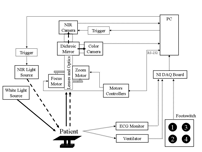
As part of this project, we have developed a novel optical imaging system that utilizes invisible near infrared light to see human disease intraoperatively. By introducing exogenous near infrared fluorescent contrast agents into the surgical field, the surgeon can perform sentinel lymph node mapping, resect tumors, and avoid critical structures, such as vessels and nerves, all under image guidance. This technology has been licensed nonexclusively to GE Healthcare for translation to the clinic and is the subject of a Bioengineering Research Partnership for the . Detailed schematics and other downloads related to this project are available below. The Frangioni Laboratory has constructed a state-of-the-art operating room in which a next generation imaging system and contrast agents are tested using the concept of spiral development. In this environment, engineers, surgeons, and chemists work together on a daily basis to improve surgical techniques. |
|
|
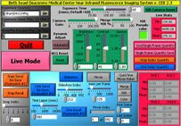
To take advantage of the numerous FDA-approved SPECT radiotracers, we have added three-dimensional radio-scintigraphy capabilities to our intraoperative NIR fluorescence imaging system.
This project is a multi-institutional collaboration among the Frangioni Laboratory of Beth Israel Deaconess Medical Center, the Lanza Laboratory at Massachusetts Institute of Technology, the Idoine Laboratory at Kenyon College, and the Accorsi Laboratory at Children’s Hospital Philadelphia. To maximize field of view, sensitivity, and resolution for tumor detection, and to minimize data acquisition time, we are utilizing coded aperture collimators and have developed a Labview VI for automated evaluation of coded aperture design and performance. |